
CELLAVISTA4 and NYONE HE4 compatibility with new industry standard of assuring monoclonality by red/green distinguishable cell studies
BioPhorum Operations Group (BPDG Monoclonality Team) has published a very important paper in PDA (Parental Drug Association) presenting their consensus approach to proving monoclonality using image based systems.
Implementation of Plate Imaging for Demonstration of Monoclonality in Biologics Manufacturing Development PDA J Pharm Sci Technol; July/August 2018 72:438-450; accepted version published online: April 18, 2018; doi:10.5731/pdajpst.2018.008789, vol. 72 no. 4 438-450, https://www.biophorum.com/monoclonality-paper-publ…
The paper gives highly detailed information about the application of imaging with regard to proving monoclonality, specifically focusing on concerns regarding single cell imaging. These concerns, namely ghost wells and false positive clonal wells are addressed and explained, as well as strategies outlined on how to mitigate the risks in terms of using the images for incorporation in cell line generation reports as part of the BLA (biological license application).
Ghost Wells (containing colonies without a single cell image) have been the specific focus of several companies who have already presented strategies on how to generate statistically relevant and robust data in several presentations at conferences as well as in peer reviewed publications.
Evans et al. (2015), Assurance of monoclonality in one round of cloning through cell sorting for single cell deposition coupled with high resolution cell imaging. Biotechnology Progress, 31: 1172–1178.; doi:10.1002/btpr.2145
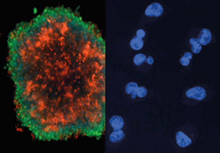
False positive clonal wells (non-imaged cell(s) dropping on other cells) have been put into focus just recently, as well as distinguishable cell (e.g. co-culturing GFP and mCHERRY expressing cells and subsequently cloning them) studies to efficiently generate robust data for a statistical analysis of this (rare) phenomenon.
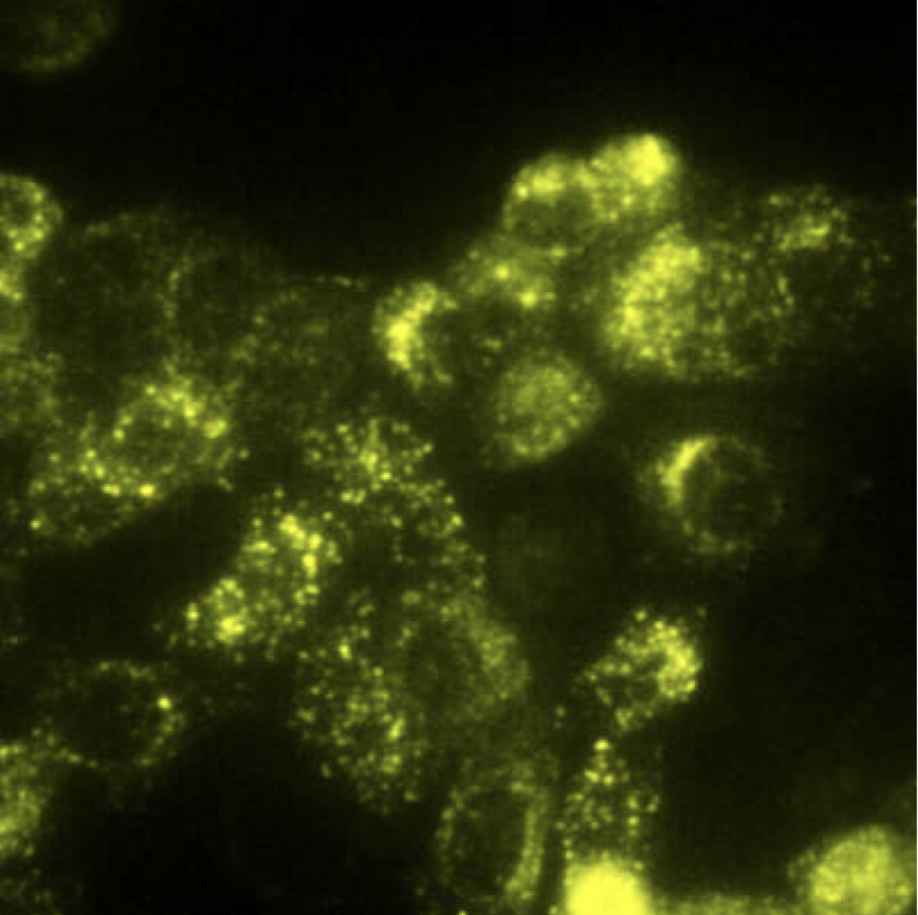
SYNENTEC’s NYONE and CELLAVISTA imagers are highly sensitive and rapid at multiplexing fluorescence imaging and are thus a perfect tool to conduct these studies. A high percentage of SYNENTEC’s installed base of imagers are already equipped with Blue/Green and Lime/Red-LP fluorescence channels to rapidly perform the measurements needed, not only in single cell stages, but also at colony level.
For this analysis, SYNENTEC specifically developed Confluence 2F (brightfield and 2 fluorescence channels) image processing tools that generate confluence growth curves for each well, and also gives insight into colony color per well and is thus the perfect image processing tool to analyze distinguishable cell studies.
Selected references:
Fieder, J., Schulz, P., Gorr, I., Bradl, H., and Wenger, T. (2017) A single-step FACS sorting strategy in conjunction with fluorescent vital dye imaging efficiently assures clonality of biopharmaceutical production cell lines. Biotechnol J 12; doi:10.1002/biot.201700002
Follit, J. (2018) Single Cell Cloning: Development, Application and Characterization of a Fluorescent Assisted Single Cell Cloning Protocol, Cell Line Development and Engineering, San Francisco
Lundquist, A. (2018) Industry perspectives and case studies towards demonstration of monoclonality for biologics manufacture development, BioProduction Congress, Dublin